


Case Study
Diagnostic Decisions Through Dental Image Analysis
Case Overview
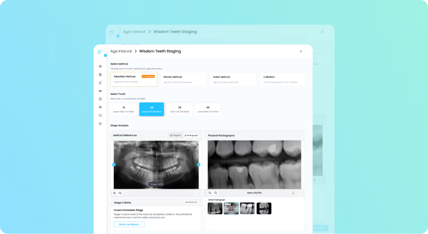
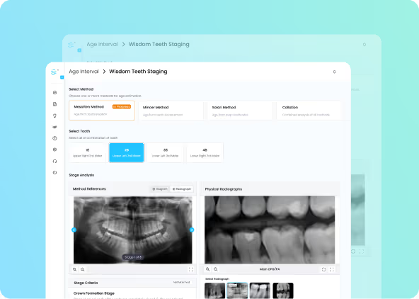
A panoramic radiograph revealed a suspected cystic lesion, prompting further diagnostic evaluation to determine the need for surgical intervention. Using the Dental ID Image Analysis module, clinicians applied polygon-based tools to precisely measure and monitor the lesion over time. The analysis revealed a reduction in cyst size, suggesting it was inactive. This radiographic evidence supported a conservative management plan, helping avoid unnecessary surgery while enhancing diagnostic confidence and demonstrating the value of advanced imaging tools in dental decision-making.

Key Findings

Lesion Assessment
Polygon-based measurement tools allowed precise tracking of cyst size and behavior over time.
Non-Invasive Technique
Image analysis confirmed regression of the lesion, supporting a conservative approach and preventing unnecessary surgical intervention.

Diagnostic Confidence
Advanced imaging tools enhanced clinical judgment by delivering measurable, evidence-based insights from radiographic data.
Medical Value & Impact

Objective Documentation
Radiographic measurements provided verifiable, time-stamped evidence suitable for inclusion in medical and legal records.

Evidence-Based Management
Clear imaging and measurable lesion changes supported appropriate treatment decisions, aligning with clinical guidelines.

Medico-Legal Reporting
Structured image analysis improved the clarity and credibility of expert documentation, supporting interdisciplinary reviews.


Case Studies
Field Applications & Case Insights
Humanitarian Support
Age verification through dental assessment
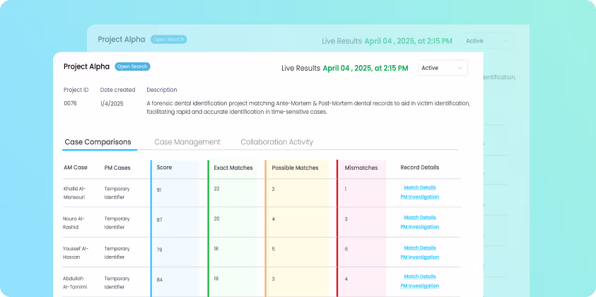
Forensic Dentistry
Victim identification through dental records
Age Verification Tool
Legal Age Verification Through Dental Assessment
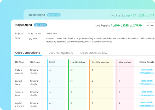
Dental Forensics
Victim Identification Using Dental Records

Empowering professionals to perform standardized dental assessments anywhere with accuracy, efficiency, and trusted results.


Copyright © 2025 Dental ID. All rights reserved.


